The Genesis of Harrington Rods: Pioneering Scoliosis Treatment in the 1960s
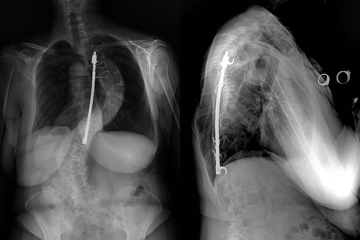
Harrington Rods were the first implants developed for the treatment of scoliosis. They were designed in the 1960s by Dr. Paul Harrington, an orthopedic spine surgeon in Houston, TX. Consisting of two hooks and a rod connecting them, Harrington rods were a massive step forward in scoliosis treatment because they allowed for significant correction of scoliosis to be achieved during the operative procedure.
Traditional Scoliosis Treatment: Methods Before Modern Surgical Advances
Prior to Harrington rods, scoliosis could be treated surgically, but was done without hardware. A surgical incision was made over the spine where the curve was present, and the spine was fused by removing the soft tissue and outer layer of the bone from the spine, and then laying bone graft over the spine to encourage the spine to heal together into one bony block. The bone graft could either come from the actual patient (autograft) or from a cadaver (allograft.) Once the fusion was done, the incision was closed, and the patient was placed in a plaster cast which was molded during its application to try to straighten the spine as the fusion healed. These patients were then typically admitted to a hospital or scoliosis ward, where they were kept on bedrest for weeks or even months as their fusion healed, in hopes of maximizing the chances of the fusion healing to prevent their scoliosis curve from worsening into the future.
Revolutionizing Scoliosis Surgery: The Introduction of Harrington Rods
Harrington rods offered a huge advancement in the treatment of scoliosis. By placing one hook at the top of the curve and a second at the bottom, and placing a rod to distract between the hooks, a scoliosis curve could be significantly straightened at the time of surgery, and allowed for much greater stability, which increased the chances of the fusion healing and allowing patients to mobilize out of bed much faster. Over time, scoliosis surgeons began implanting more than one rod, with a “distraction rod” placed on the concavity (or inside) of the curve, and a “compression rod” along the convexity (or outside), to allow for even better correction and improved stability, offering even better chances of successful fusion and allowing for earlier mobilization and faster recovery.
Advancements and Innovations in Scoliosis Surgery with Harrington Rods
Harrington rods were utilized in scoliosis and spine surgery until the 1980s when a newer system was developed in France by Dr. Cotrel and Dr. Dubousset. Cotrel-Dubousset instrumentation built on Harrington rods by offering multiple different hooks which could attach to different parts of the spine and were connected by rods, typically two rods, placed on the left and right sides of the spine. By using multiple hooks and two rods, CD instrumentation allowed for even better curve correction and better post-op stability, both allowing for improved outcomes and earlier return to normal function. In the early 2000s, Pedicle screws became popularized for the treatment of scoliosis, placing screws in the spine to get better fixation and improved correction of scoliosis curves. Pedicle screw constructs remain the standard today for spine fusion treatment of scoliosis.
The Evolution of Scoliosis Treatment: From Harrington to Cotrel-Dubousset and Pedicle Screws
While Harrington rods offered a huge advancement in the treatment of scoliosis, there are risks associated with Harrington rods, and patients who had Harrington rods implanted as children or young adults frequently develop further issues into adulthood. Flatback Syndrome is a well-documented complication of Harrington rod placement. While the spine is supposed to be straight when viewed from the front or back, from the side, the spine is supposed to have curves including thoracic kyphosis and lumbar lordosis. These curves develop during infancy and early childhood, and they are balanced to allow for the head to be aligned with the pelvis when standing up straight. Harrington rods worked by distraction, meaning the scoliosis curve was straightened by placing a hook at the top of the curve and a hook at the bottom, a rod was placed between the hooks along the concavity or inside of the curve, and the hooks were distracted along the rod in an attempt to straighten the curve. If the curve extended into the lumbar spine, and the distraction occurred in the lumbar spine, this distraction counteracted against the normal lumbar lordosis curve (seen in the side view) which would make the lumbar spine straight when viewing it from the side. This lumbar straightening would result in Flatback Syndrome, where over time patients develop worsening posture and inability to stand up straight, particularly as the discs and motion segments below the Harrington rod would wear out, causing progressive worsening Flatback Syndrome.
The Complications of Harrington Rods: A Closer Look at Flatback Syndrome and Progression of Scoliosis
Patient’s with Flatback Syndrome frequently require revision surgery, with the goal to decompress the nerves that are being pinched or compressed, and to reconstruct the spine so that the patient can stand up straight. Sometimes this can be achieved by extending the fusion distally one or more segments. A Pedicle Subtraction Osteotomy (or PSO) is a procedure that also allows for correction of Flatback Syndrome by correcting the flatback by re-creating the curvature that was lost through a previously fused area of the spine. Progression of Scoliosis is another risk associated with Harrington rods. While Harrington rods offered significant improvement in scoliosis correction over non-instrumented efforts, they were not perfect, and significantly less effective than current correction techniques utilizing screws and multiple rods. Scoliosis curves were frequently under-corrected, and there was a higher risk of developing a pseudarthrosis or non-union, meaning the spine wouldn’t fully heal together into a bony block. Under-corrected curves are at higher risk of worsening scoliosis above or below the surgically treated area. Pseudarthrosis and non-union of fusion also increase the risk of progressive worsening scoliosis at the operated curve. Pseudarthrosis also increases the risk of pain over the curve where the spine doesn’t heal properly. The patients frequently notice pain or worsening curvature over time after their Harrington rod surgery, and surgical treatment for revision of the Harrington rod may be necessary to address a worsening deformity or increasing back or leg pain or worsening neurological symptoms.
Modern Solutions for Harrington Rod Complications: From Revision Surgery to Advanced Techniques
Many of these patients also develop progressive worsening back pain and lower extremity pain, numbness, or weakness. In these cases, revision scoliosis surgery offers the potential to correct spinal deformity and improve clinical symptoms. If you have had a Harrington rod surgery, and are concerned about potential worsening scoliosis or painful symptoms, please contact Dr. Jason Lowenstein.
