Vertebral Column Resection
What is a Vertebral Column Resection?
A Vertebral Column Resection (VCR) is a type of osteotomy that is used to correct severe cases of spinal deformity. Osteotomy simply means that your doctor will remove bone. However, a VCR takes this one step further.
During a VCR, your doctor will remove or “resect” one or more vertebrae from your spine. A fancier phrase for your spine is “vertebral column.” This refers to the fact that the vertebrae in your spine are stacked like a pillar. Hence, the term: Vertebral Column Resection.
Milder forms of osteotomy do not remove entire vertebrae. These include the Posterior Column Osteotomy (PCO) and the Pedicle Subtraction Osteotomy (PSO). Instead, these procedures resect tiny sections of bone on the back side of your spine.
However, if you have >100 degrees of curvature, then removing small sections of bone just won’t cut it. You’ll need a VCR to fix your spinal alignment.
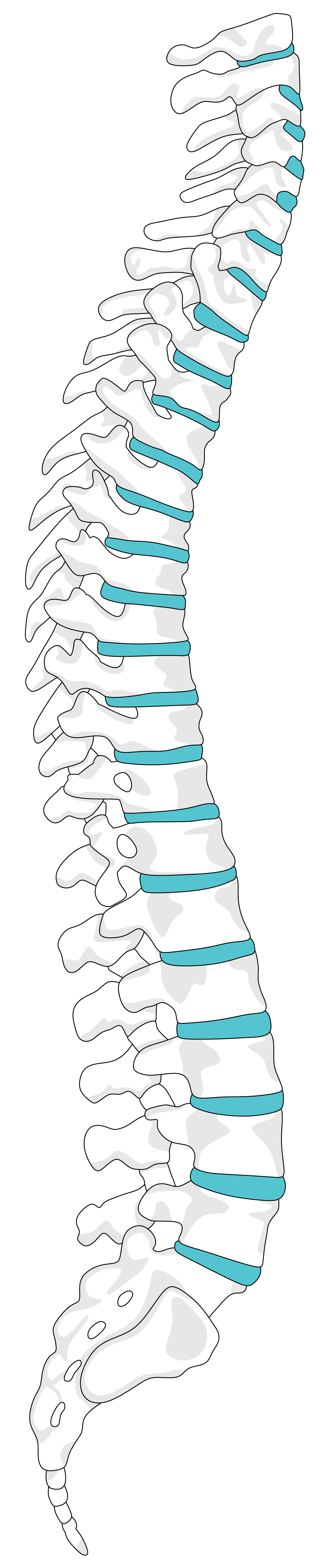
But first, let’s revisit a “healthy” spine. A healthy spine isn’t ramrod straight. Instead, it contains gentle inward arcs (known as lordosis) and subtle forward curves (known as kyphosis). If too much kyphosis occurs, the shoulders round and the head pitches forward. You may hear of this referred to as a sagittal imbalance because the head is no longer balanced over the pelvis.
When viewed from the front, however, the spine should seem straight. If the spine juts outward to the side, scoliosis (a form of coronal imbalance) occurs.
Severe scoliosis or kyphosis (>100 degrees) can cause pain, lung and heart problems, issues with walking, and loss of confidence. A VCR can permanently reverse these symptoms.
How is a Vertebral Column Resection Performed?
If you need a VCR, your surgeon will position you face-down on a Jackson frame. This operating table contains a gentle slope that exposes the back side of your spine.
Depending on your situation, your doctor will enter your back from a posterior (back) or anterior (front) approach. Sometimes, a surgeon can achieve best results by using a combined, or anterior and posterior, approach. However, entry from the back is the most common.
Next, your surgeon will fasten pedicle screws to your vertebrae. These will attach to the vertebrae above and below the resection area. Then, your surgeon will remove the back portion of the targeted vertebrae. This will include your:
- Spinous Process: The three bony points on the back side of your vertebrae
- Lamina: The bony shell that guards your spinal cord
- Facet Joints: The joint where two vertebrae come together
- Pedicles: The swatch of bone that joins the vertebral body to the processes
With these items removed, your surgeon can now see your spinal cord and the round front half of your vertebra, known as the vertebral body. After removing the left side of the vertebral body, your surgeon will add rods to each screw. This crucial step in the surgical procedure protects your fragile spinal cord, which is now exposed.
Finally, the problematic vertebra can be excised. If more than one resection is needed, your surgeon will repeat the steps above. In addition, your surgeon may remove ribs, if your thoracic spine is the target of the procedure. With all vertebrae removed, a wedge-shaped gap will remain in the back side of the spine. Your doctor will then realign the spine and close this gap. Closing this hinge folds the spine backward onto itself and into a more ideal shape.
Will I Need Spinal Fusion with a Vertebral Column Resection?
Yes. Your surgeon will use a combination of implants and bone grafts to support your spine as it heals.
Specifically, graft material can be sourced from your hip or a donor supply. If needed, your surgeon may even opt to use a cage–a metal device filled with bone graft material. As the bone tissue matures, it will grow through the “bars” of the cage to fuse to your spine.
In addition, your doctor will add extra rods to brace your spine.
The ultimate goal of these last steps is to create a spine that is stable, durable, and pain-free. With the final pieces of hardware in place, the osteotomy procedure is now complete.
Do I Qualify for an Anterior Lumbar Interbody Fusion?
You may need an ALIF if you have:
- Scoliosis: If you have less than 40 degrees of scoliosis and your condition did not improve with bracing. This method can be used for all types of scoliosis, including pediatric scoliosis.
KyphosisIf you have less than 40 degrees of kyphosis. This includes kyphosis that results from degenerative disc disease. (Note: Your surgeon may perform an ALIF in conjunction with an osteotomy to correct your kyphosis). - Lordosis: If you have less than 40 degrees of swayback, aka hyperlordosis.
- Spondylolisthesis: If you have a vertebra that has slipped more than 50% forward over the vertebra beneath it.
- Disc Problems: If your spinal deformity caused the discs in your back to bulge or rupture.
Unfortunately, an ALIF isn’t for everyone. You may not qualify if you have or have had:
- Severe osteoporosis or weak bones.
- Compression fractures
- Spinal Tumors
- Scarring from previous abdominal surgeries
To find out if you qualify for an ALIF, contact Dr. Jason Lowenstein, MD today! Dr. Lowenstein is a fellowship-trained scoliosis surgeon with over a decade of surgical expertise. Because you deserve only the best, contact one of NJ’s top-rated spine surgeons today!