Spinal Alignment Issues: Scoliosis, Kyphosis, & Spinal Deformity
In this filmed lecture, Dr. Jason Lowenstein, Director of Scoliosis and Spinal Deformity at Morristown Medical Center, delivers a lecture on spinal deformities with a particular focus on scoliosis diagnosis and treatment. Dr. Lowenstein, founding member of The Advanced Spine Center, uses his spinal deformity expertise to educate patients and practitioners alike.
Are you concerned that your child (or even yourself) may be suffering from a spinal deformity such as scoliosis, kyphosis, or spondylolisthesis? If so, review the following video lecture to learn how you can spot the warning signs of these common pediatric and adult spine conditions. Then, contact The Advanced Spine Center to set up a medical consultation. You or your child may qualify for conservative treatments, such as spinal bracing or physical therapy. To find out more, contact our devoted team of patient advocates today!
Video Transcript:
I’m Jason Lowenstein. I’m a scoliosis and spinal deformity surgeon and I’m the Director of Scoliosis here at Morristown. I’m going to talk to you today about scoliosis, spinal deformities, and spinal imbalance.
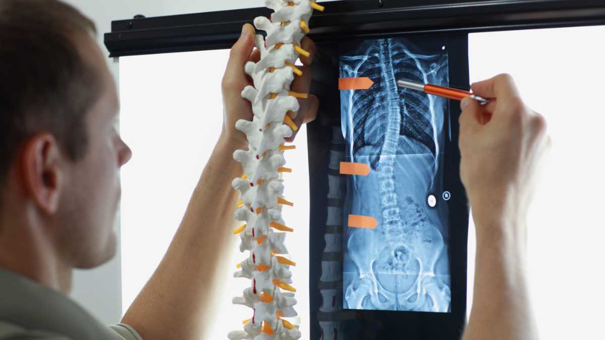
So, a spinal deformity is really considered any imbalance or malalignment of the spine. It can happen to children. It can happen to adults. And, it can really happen to any portion of the spine. When you look at the spine, it’s supposed to look straight when you look at it from the front and it’s supposed to have these curves—thoracic kyphosis, lumbar lordosis—which balance the spine and keep your head directly above your pelvis.
This patient is obviously imbalanced. I was trying to give you a really subtle case of scoliosis.
And, so typically, when you stand up straight, your head is directly above your pelvis from the front and from the side. That’s a pretty stable and controlled environment. When you start leaning forward and can’t stand up straight, you start getting pain. As well as if you stand to the side… if you have a spinal deformity which causes you to shift to the side, that can also cause pain.
So, the French. and particularly Dr. Dubousset. sort of described this idea of a cone of economy, which basically means that if you’re standing up straight (your head is directly above the pelvis when you look at yourself from the front or the side), you have to expend very little energy to be in that position.
Every degree off of that main axis, if you start leaning forward or to the side or backward, and you have to stay fixed in that position, you have to expend more energy to stay in that position. Over time, your muscles fatigue. You start having pain. And, eventually, you need an assistive device like a cane, a walker, a wheelchair… in order to be able to stand up straight.
So, this is an example of that kind of patient, right? This patient, this cone of economy, is off to the right side but also leaning forward. If you draw a line from their head down, they are in front of their pelvis. So, these patients tend to develop progressive pain.
When I see a patient in the office, the first thing I want to do is take a history to try to figure out: “Do they have a deformity? And, if so, where is this deformity coming from?”
In this particular patient, you see a bunch of stuff. You see that they have a crease right here, which shows asymmetry. You see that this shoulder blade is higher than this one. You’re looking for that asymmetry. You really have to actually have the patient put on a gown and look at their back. Sometimes, you have to look at their front. You have to see if they have any abnormalities in their chest wall as well.
Here’s a couple of different patients. These are young adult patients. This patient obviously has a shoulder asymmetry. This patient has a significant trunkal shift. Their whole body is shifting forward. You can see their scoliosis. This patient is leaning forward. His ribs are elevated. If you can’t see that, this is a really obvious one. And, there’s this picture. When you see a patient with scoliosis, typically, they are supposed to have a normal alignment when you look at them from the side. If they don’t, then they have something else going on.
So, this is a patient that has curvature. That’s not this patient. This patient has Scheuermann’s Kyphosis. They have curvature. They have wedging in multiple vertebral bodies that results in this spinal deformity.
Here’s another patient with a bulge in their lower back. This looks prominent, right? So, this patient has a spondylolisthesis. What you’re seeing is the spinous process of L5, because that’s staying there and the rest of the spine is shifting forward. So, these subtle cues can let you know that there’s a spinal deformity that they have to deal with.
We also want to do a neurological examination. A lot of times, patients with a spinal deformity have a simple, straight forward scoliosis with no significant neurological changes. But, particularly in adult patients, you can start to see weakness because as they develop scoliosis, they start getting spinal stenosis. My colleagues all discuss that. They start getting narrowing of the space available for the nerve roots. They may start having radicular symptoms, such as pain going down their legs. They may have weakness or abnormal reflexes or sensations along with those changes.
And, then we get x-rays. And, typically you want to get a 3-foot standing scoliosis x-ray, which is an x-ray of the entire spine. It shows the neck. It shows the shoulders. It shows the spine. It shows the pelvis. Because you’re looking at the entire spine as well as the alignment of the spine in comparison to where their head is relative to their pelvis.
So, we measure something called the Cobb angle. Cobb angle is a way to describe how severe their curve is and what the degree of magnitude is. There’s a couple of different reasons. One, you’re going to do this so you can talk to your colleagues, explain what their deformity is. But, you do this also if you’re watching a curve progress over time. You can say “Okay, at this point in time it measured this.” And, if it progressively worsens, sometimes this is an indication that they need to have more treatment.
When do we order advanced imaging? It really depends and it’s on a case-by-case basis. If a patient does have an abnormal neurological examination, oftentimes we’ll get an MRI. The reason why is we’re looking to see if something is going on in the spinal cord, which would be both allowing scoliosis to occur in the first place and potentially making it worse.
This right here is a patient with left thoracolumbar scoliosis. But, this right here is a syrinx. It’s a collection of fluid in the center of the spinal cord that’s not supposed to be there. That can cause scoliosis but also contribute to scoliosis progressing over time.
So, when do we get advanced studies like a CT or myelogram? You know, it’s becoming less and less common to get those. But, in patients who have stainless steel hardware, particularly in patients who had surgeries in the 80s and 90s, you can’t see very well on an MRI. So, we’ll get a myelogram. For anyone who doesn’t know what a myelogram is, the radiologist will inject dye into the nerves, into the dural sac around the nerves. The dye will flow up and down. You start by getting x-rays. That’s the myelogram.
You can see here is the dye flowing up. It stops here. It stops there. This may give us an indication that the patient has spinal stenosis at those levels. Then, you get a post-myelogram CAT scan. Severe stenosis here. Severe stenosis here. And, at L5 and L5-S1 where the patient had previous surgery, it looks like it’s wide open.
Okay, so what are the treatment options for scoliosis and spinal deformity? There’s a lot of stuff you can do and we usually try to start out conservatively. So, you can give medications. And, I think we already discussed a lot of medications that you can give. You can try bracing. You can try chiropractic treatment. You can try physical therapy. You can try pain management, including steroid injections. And, eventually, you can go on to have surgery.
People oftentimes ask about bracing for scoliosis because they had a friend in middle school or high school who needed to wear a brace. It’s very typical to brace children who have scoliosis. The reason why is that the spine is still forming and modulating while you are young. Before you obtain skeletal maturity, the bones are basically made of cartilage and we can affect the way they grow by bracing. The idea behind bracing is that you put a patient in a brace, you put a corrective force on the brace, and you’re trying to straighten the spine in the brace to prevent the curve from progressing over time so they don’t need to have surgery.
In adults, it’s a little bit different because the spine has already stopped growing. So, bracing doesn’t offer the same effect.
So, here’s a picture of different braces. Typically, the idea behind bracing is that if you’re going to wear a brace, you really need to wear it as much as possible in order to make it work. I like this picture because these are two children wearing braces. These are actually both advertisements to try to sell braces. This is a flexible brace. It doesn’t really work, but she looks fairly happy. This is a brace that’s called the Milwaukee brace, which was back in the 60s and 70s. This was the happiest kid that they could find to put in this brace and take a picture. You know, it’s clearly not super successful.
In terms of bracing, we do have data to support that bracing works in kids. And, not to go too off the chart on this, but there’s a guy called Stu Weinstein who did a study at the University of Iowa. It was a multi-center study. It was an NIH-funded study. For those of you guys who do care about peer-reviewed studies, it’s hard to receive NIH funding for stuff. You have to have lots of patients and lots of (statistical) power. NIH pulled the study after two years because they said the data was so overwhelmingly supportive that braces work that by not bracing kids with scoliosis we were doing a general disservice to the public.
So, what they did, they did a randomized study where they either had kids wear a brace or not wear a brace. They were skeletally immature. They wanted to see if the curve progressed or did not progress. There was a high correlation between not wearing a brace and having the scoliosis progress.