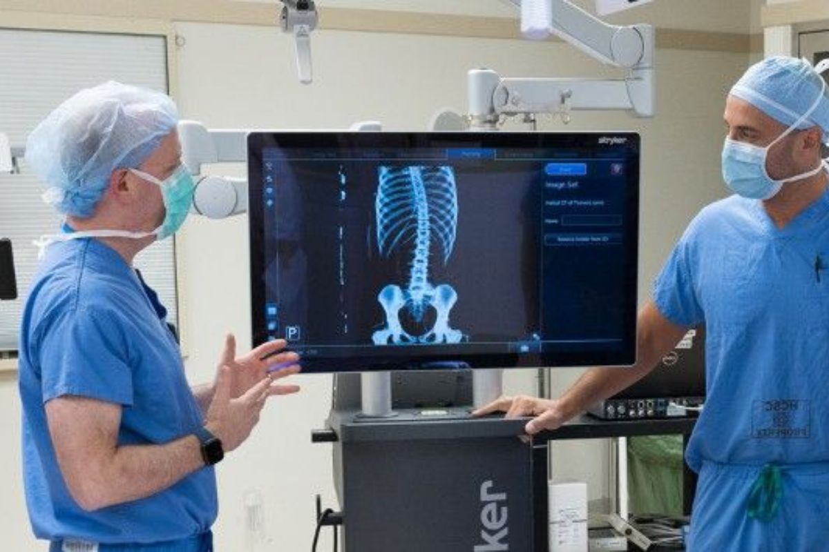
Advanced Imaging for Complex Scoliosis Correction
Surgeons at the Scoliosis and Spinal Deformity Center at Morristown Medical Center are making scoliosis and back pain treatment easier with technology that enables them to essentially see through the skin into the patient’s spinal canal.
Scoliosis is an abnormal lateral curvature of the spine. It is most often diagnosed in childhood or early adolescence. If left untreated, over time scoliosis can cause pain and various other health problems. Scoliosis affects six to nine million Americans.
Most cases of scoliosis are mild, but some curves worsen as children grow. Severe scoliosis can be disabling. An especially severe spinal curve can reduce the amount of space within the chest, making it difficult for the lungs to function properly. In these cases, surgical correction is usually the recommended course of treatment.
When used with Stryker’s navigation platform, Airo helps allow for screws to be placed in the spine with pinpoint accuracy, providing an opportunity to improve both speed and safety during complex scoliosis and spinal deformity correction. Jason E. Lowenstein, MD, Director of Scoliosis and Spinal Deformity at Morristown Medical Center
A New Approach to Treating Severe Scoliosis
For patients with severe scoliosis, for which surgical correction is necessary, surgeons at the Scoliosis and Spinal Deformity Center were the first in the nation to use a new type of portable CT scanner to guide their surgery and actually “see” inside the spinal canal during surgery.
The procedure is done minimally invasively, placing a tiny thorascopic camera into the chest through a small incision to visualize the spine on a video monitor, and then using Stryker’s Airo TruCT Mobile Imaging system to obtain a CT scan of the patient during the surgery. Using these images with Stryker’s navigation software, as well as direct thorascopic camera visualization, spinal screws can be placed into the spine using real-time CT guided navigation. This allows the screws to be safely and accurately placed into the spine.
The use of advanced intraoperative imaging helps enable surgeons to perform the least invasive surgical correction possible for the patient, which can result in less postoperative pain and a quicker recovery time when compared to more conventional treatments.
Benefits of Stryker’s Airo TruCT Mobile Imaging
- Provides intuitive visual access to difficult-to-reach spinal canal
- Airo TruCT can give surgeons real time pre- and post-operative knowledge in the operating room
Benefits of Minimally Invasive Spinal Surgery
- Potential for less blood loss, less chance of infection and shorter recovery times
- Potential for shorter hospitalizations than traditional open surgery
Conditions We Can Image With Airo TruCT
- Degenerative disc disease
- Herniated disc
- Sciatica
- Scoliosis (pediatric and adult)
- Spinal deformities (e.g., kyphosis, scoliosis, etc.)
- Spinal stenosis
- Spinal trauma
- Spinal tumors
- Spondylolisthesis
